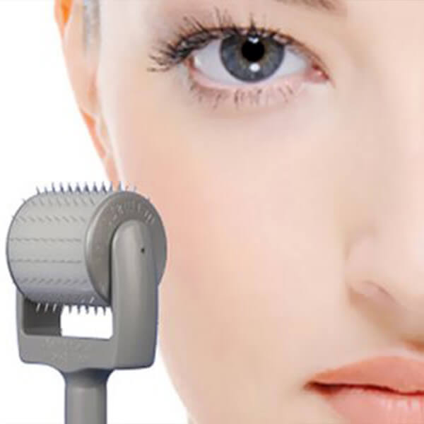
Skin needling involves puncturing the dermis with a needle-like device to induce new collagen for the purpose of skin rejuvenation. This technique involves a controlled injury to blood vessels, which involves a number of complex cellular changes. Blood platelets spill into the injury site, causing the platelets to come into contact with the extracellular matrix. This contact triggers the platelets to release clotting factors, essential growth factors and cytokines, such as Platelet Derived Growth Factors (PDGF) and Transforming Growth Factor Beta (TGF beta). Once the bleeding has ceased, neutrophils enter the injury site and begin phagocytosis to remove foreign material, bacteria and damaged tissue. Macrophages also enter the injury site and continue phagocytosis and release more PDGF and TGF Beta.
Once the injury site is cleaned, Fibroblasts migrate into the injury site a start to produce and deposit new extracellular matrix. The new collagen matrix becomes cross-linked and organized during the final remodeling phase, which can continue for up to 12 months after injury has occurred. Collagenesis is a process that naturally promotes collagen production in your skin without damaging it. Collagen Type III is the dominant form in the early wound-healing phase. Collagen III is gradually replaced by collagen type I over a period of 12 months. Needle holes are about 4 cells wide, while the epidermis and dermis remain intact. The fact that the epidermis stay intact is extremely advantageous as it means less downtime and potential post inflammatory hyper pigmentation is minimal.
